Congratulations to our Nano Image Winners of the “Plenty of Beauty at the Bottom” 2019 NSF NNCI Contest
In the “Most Stunning” category…

In the “Most Unique Capability” category……..

In the “Most Whimsical” category…

Thank you to all contestants of the 2019 KY Multi-scale Nano Image Contest!!!

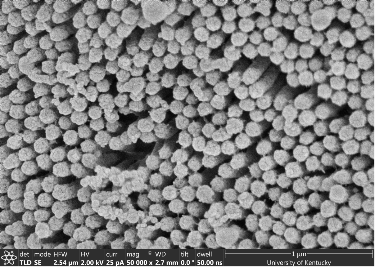
Image by Ahamed Ullah
Image title: SEM Image of Cr Doped MnO2
Image description: Cr-doped MnO2 was synthesized by hydrothermal method. With the doping of Cr the morphology of the pure MnO2 (nanowire) changes to “Urchin” like shape for Cr-doped MnO2. This material is a precursor for the synthesis different Na based cathode material for Na ion battery.

Image by: Jasmin Beharic
Image Title: Petrified Wood
Image description: SEM Image taken at the Micro Nanotechnology Center, University of Louisville. Petrified wood is fossilized remains of vegetation that has transitions to stone over thousands of years. This particular piece of petrified wood was imaged for the local artist "Matt Weir" who is working on a art exhibition based around this piece of petrified wood. He is trying to show how the same object can be interpreted in many different ways.

Image by Emtias Chowdhury
Image title: Ultra Low Calorie Nano Cake Pops
Image description: Anisotropic Nanoprism Coated Polymer Beads. DNA mediated interaction between nanoscale gold nnaoprisms and microscale polymer beads. Emtias Chowdhury is a Grad Teaching Assistant from A&S Chemistry, University of Louisville.

Image by Sanju Gupta
Image title: The Entrance to Lost River Cave, Bowling Green, KY.
Image description: Boron nitride nanosheets (BNNS) and poly(vinyl) alcohol 2wt.% are mixed, ultrasonicated and processed in liquid nitrogen ‘ice templating’ followed by freeze-drying at -40 oC to construct three-dimensional directed assembly composites. The layered morphology along with polymer strands connecting the BNNS sheets is apparent in falsely colored scanning electron microscopy image acquired at EMC. Credited to Dr. Sanju Gupta and Alex Henson, Department of Physics & Astronomy, Western Kentucky University, Bowling Green, KY.

Image by: Brian Wadjdyk
Image title: “It’s an SEM photo finish...Nano-Secretariat wins by 3 voxels!”
Image description: The Nano Horse structure was fabricated at the KY Multiscale NNCI Site using Nanoscribe 3D printing technology. Nano Jockey hand-drawn by Fine Arts undergrad Mallory Lucas.

“Nano Bullet”
Author: Chuang Qu
NNCI KY Multiscale Core: Huson Imaging and Characterization Lab, MNTC, UofL
Tool: Supra SEM
Description: Germanium nanobullet created by Glancing Angle Deposition (GLAD) using electron-beam evaporator in the UofL cleanroom.

Image by: Maria Kosmidou
Image title: Nanoporous Niobium
NNCI KY Multiscale Core: Electron Microscopy Center, UK
Tool: FEI Helios Nanolab 660
Image description: Cross section of Nanoporous Niobium thin film (approx. 1 micron of thickness), fabricated after Vacuum Thermal Dealloying Technique from a Magnesium based precursor alloy (at. % Mg75Nb25) via Physical Vapor Deposition. Figure contains three frames in different magnifications indicating both the nanoscale (higher magnification) and the microscale porosity (lower magnification). For the sake of homogeineity, a thin dense layer of Niobium has benn deposited on the top of the nanoporous film.

Image by: Md Emtias Chowdhury
Image Title: Gold Nanoprisms

Image by: Md Uddin
Image description: SEM 4300 images of Ag nanowires. These nanomaterials were synthesized for catalytic ad thermoelectric applications.

Image by: Md Uddin

Image by: Md Uddin

Image by Chuang Qu
Image title: We Serve Fresh Nano Rotini!
Image description:The germanium rotini we are serving is made in an electron-beam evaporator. In this high-vacuum environment, germanium is heated up by electrons, evaporated, and deposited slowly onto the ‘plates’. The rotini shape is formed by meticulously maneuvering the 'plate'. The rotini is ~1800 nm long; the thinnest part of the rotini is ~80 nm, which is 1/1000 the size of human hair! Have a ‘small’ bite and let me cook know which flavor you like the best! The nano-rotini is ‘cooked’ and characterized at Micro/Nano Technology Center (MNTC), Kentucky Multi-scale Manufacturing and NanoIntegration Node (KY MMNIN).

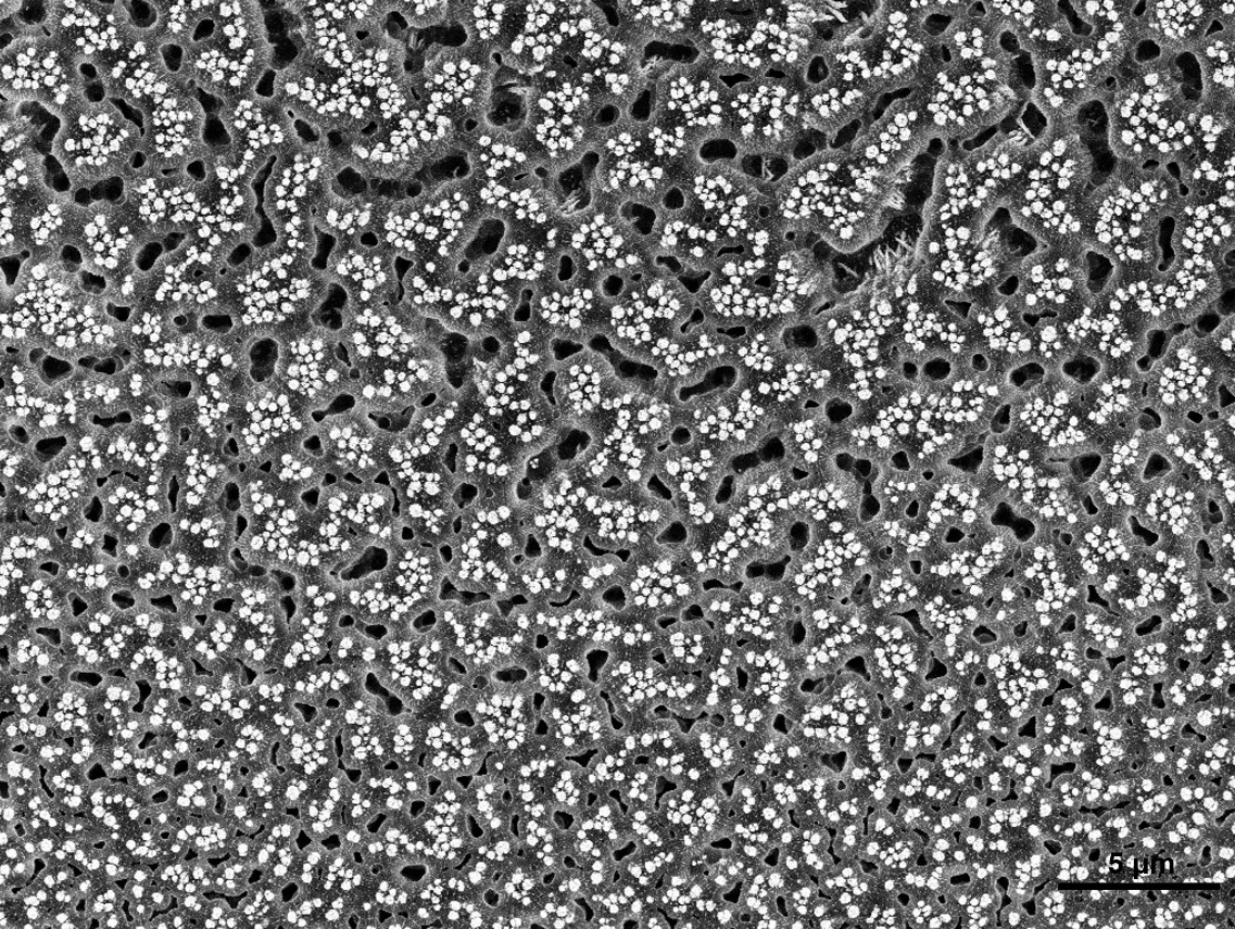
“Mindful of Microvilli”
Author: Sarah Elefson
NNCI KY Multiscale Core: Electron Microscopy Center, UK
Tool: FEI Helios Nanolab 660
Description: This image of microvilli, located at 75% of the total length of the small intestine in a 21 day old pig, expands knowledge about development and function of the small intestine. Focusing on changes of the microvilli, which are responsible for nutrient absorption and resulting growth, using micrographs produces a visual aid that shows physical changes during critical stages of life. Micrographs provide details of morphological changes during gut development and allows for evaluation of alternate diets or management at specific times (e.g., immediately following birth and/or weaning) to improve gut health and nutrient utilization in pigs.

Image by Mansoor Sultan
Image title: United We Stand, Divided We Fall.
Image description: This image was taken at CeNSE at the university of Kentucky. It was a 3D printed multi-layered color filters. Fisher-scientific environmental scanning electron microscope was used to take the image. The image provide an insight to the abilities of two-photon lithography system (Nanoscribe) at CeNSE.
“Mindful of Microvilli”
Author: Sarah Elefson
NNCI KY Multiscale Core: Electron Microscopy Center, UK
Tool: FEI Helios Nanolab 660
Description: This image of microvilli, located at 50% of the total length of the small intestine in a pig 2 weeks post-weaning, allowed for measurements of microvilli dimensions. Subsequent calculation of microvilli dimensions, results in the amazing realization that total absorptive area is increased 120,000,000X compared to absorbing from a simple tube. Assessing the development of microvilli allows for increased understanding of nutrient absorption during a pig’s life. Micrographs provide details of morphological changes during gut development and allows for evaluation of alternate diets or management (e.g., immediately following birth and/or weaning) to improve gut health and nutrient utilization in pigs.

Image by: Md Uddin
Image title: Growing of Herbs on Dead Tree Logs
Image description: SEM 4300 image of PbS NWs with extra art for 3rd category. This image shows 'Growing of herbs on dead tree logs'.

“Nano Sunflower”
Author: Chuang Qu
NNCI KY Multiscale Core: Huson Imaging and Characterization Lab, MNTC, UofL
Tool: Supra SEM
Description:
A major plant grown on the island of Lilliput. The seeds are less than 50 nanometers, but they feed the Lilliputians really well. The Nano sunflower seeds are made of Germanium by Glancing Angle Deposition (GLAD) using electron-beam evaporator in the UofL cleanroom.

Image by: Maria Kosmidou
NNCI KY Multiscale Core: Electron Microscopy Center, UK
Tool: FEI Talos F200X
Image description: Cross section of Nanoporous Niobium thin film (approx. 100nm of thickness), fabricated after Vacuum Thermal Dealloying Technique from a Magnesium based precursor alloy (at. % Mg75Nb25) via Physical Vapor Deposition. The lift-out specimen has been prepared via Focused Ion Beam. Figure contains five frames: A High-Angle Annular Dark-Field imaging (HAADF), which is a STEM technique that is highly sensitive to variations in the atomic number of atoms in the sample, as well as four frames with EDSX results of Niobium, retained Magnesium, formed Oxygen and Silicon substrate (as shown from left to right).
“Labyrinth”
Author: Chuang Qu
NNCI KY Multiscale Core: Huson Imaging & Characterization Lab (HICL)
Tool: Supra SEM
Description: Germanium nanopillars created by Glancing Angle Deposition (GLAD) are embedded into a parylene layer.
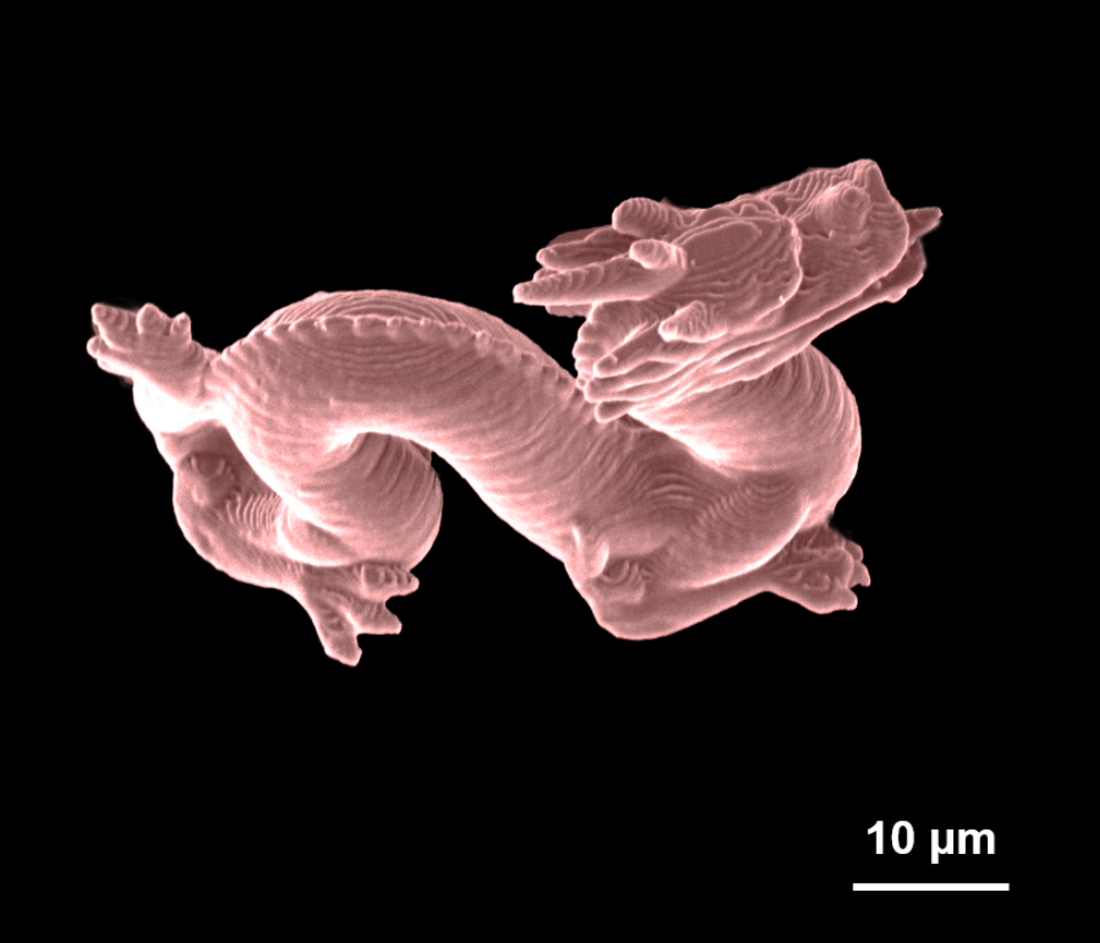
Image by: Sarah Lami

“Nano Mushroom”
Author: Chuang Qu
NNCI KY Multiscale Core: Huson Imaging & Characterization Lab (HICL)
Tool: Supra SEM
Description: Another plant grown on the island of Lilliput. The Nano mushrooms are made of several layers of Germanium by Glancing Angle Deposition (GLAD) using electron-beam evaporator in the UofL cleanroom.

Image by: Sarah Lami

Image by: Md Emtias Chowdhury
Image title: MOF Nanoparticle

Image by James Hower
Image title: The Neodymium Cat
Image description: The image of calcium phosphate mineral (florencite) in the Manchester coal, Clay County, Kentucky turns into a an abstract picture of a cat when the neodymium in the mineral and the silicon in the surrounding matrix are emphasized. The High-angle annular dark-field scanning transmission electron microscopy (HAADF-STEM) image was produced as part of a study of the sources of rare earth elements in coal.

Image by Ahamed Ullah
Image title: Cr Doped NaxMnO2
Image description: Cr-doped NaxMnO2 was synthesized by conventional solid-state method by annealing the mixture of Cr-doped MnO2 and Na2CO3. This cool and lattice plane resolved image of Cr-doped NaxMnO2 nanowires is an indication of the single-crystallinity of this nanowire. This material is used to study the Na-ion migration through the material using high resolution TEM by applying the electrical bias in the real time.

Image by JIe Ma
Image title:
Image description: The STEM (scanning/transmission electron microscopy) image and element maps of NaGdF4,20%Yb,2%Er nanoflowers made of nanoneedles prepared by a Pulsed Laser Irradiation in Solution synthesis strategy.

Image by: Ahamed Ullah
Image title: SEM Image of W Doped VO2
Image description: Single-crystalline W-doped VO2 was synthesized by hydrothermal method. The metal to insulator transition (MIT) of this material as a function of electrical bias is studied using in situ TEM in the real-time.

Image by Ning Wei
Image title:
Image description: This image is the EDS color mapping of silver nanoparticles supported on zeolite Y. Zeolite Y is a porous material consists of SiO2 and Al2O3. In this image, each element is in a different color. Ag nanoparticles with a particle size of 5-10 nm are on the surface of zeolite Y. Silver nanoparticles supported on zeolite Y is a catalyst that can catalyze the oxidation of phenolic structures either with light or heat. Now, this catalyst is used to treat lignin, the second most abundant biomass in the world, to yield valuable chemicals.

Image by Sarah K Lami
Image title: “Moon City in the Nano Universe”
Image description: Moon City's sculpture was fabricated at the KY Multiscale NNCI Site using Nanoscribe 3D printing technology.
”Shoot for the moon. Even if you miss, you’ll land among the stars.” – Les Brown

Image by: Maria Kosmidou
NNCI KY Multiscale Core: Electron Microscopy Center, UK
Tool: FEI Talos F200X
Image description: 3D reconstrunction of Nanoporous Niobium thin film, fabricated after Vacuum Thermal Dealloying Technique from a Magnesium based precursor alloy (at. % Mg75Nb25) via Physical Vapor Deposition. Figure contains four frames: A xy (top left) and random xyz plan view (top right) of STEM image and EDS results of Niobium, retained Magnesium, formed Oxygen and Silicon substrate for xy view (bottom left) and random xyz view(bottom right).

Image by: Md Emtias Chowdhury
Image title: Janus

Image by: Md Uddin

Image by: Md Uddin
Image by: Md Uddin
Image description: SEM 4300 images of Ag nanospheres. These nanomaterials were synthesized for catalytic ad thermoelectric applications.